Improve your research with fluorescence live-cell imaging
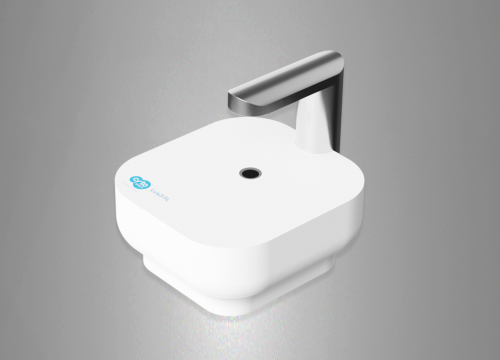
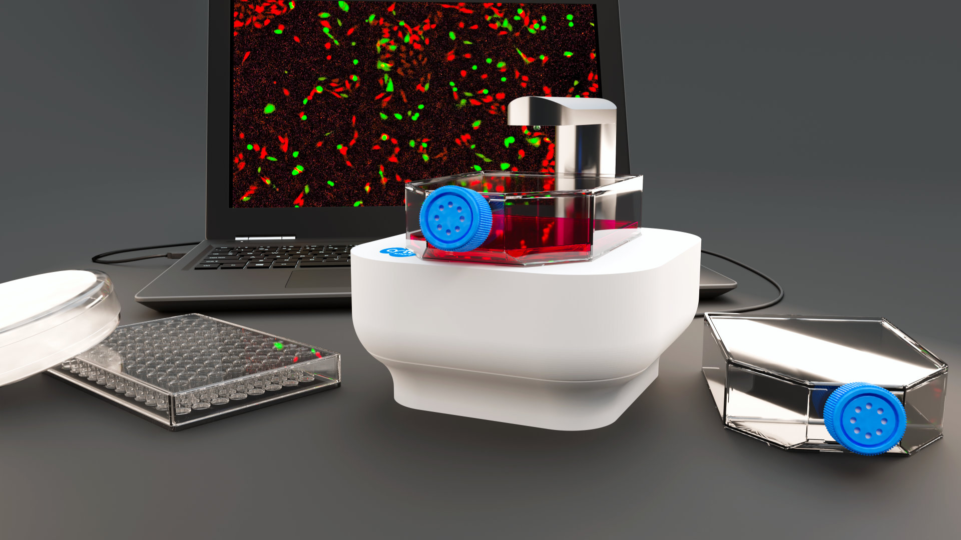
Using fluorescence live-cell imaging, researchers can get more relevant data from their experiments. The CytoSMART Lux3 FL is a small fluorescence live-cell imager equipped with one brightfield and two fluorescent channels (green and red). The device enables researchers to unravel cellular processes in real time, providing lots of valuable data about the experiments.
The CytoSMART Lux3 FL offers:
- >> Integrated image analysis of brightfield and fluorescent areas or fluorescent object count
- >> Time lapse movies to investigate the development of cellular processes
- >> Improved research with expanded number of variables for analysis
- >> Full remote access allows to inspect cell cultures without entering the lab
- >> Small, portable and easy-to-use device
Analyze cells in their desired culture environment
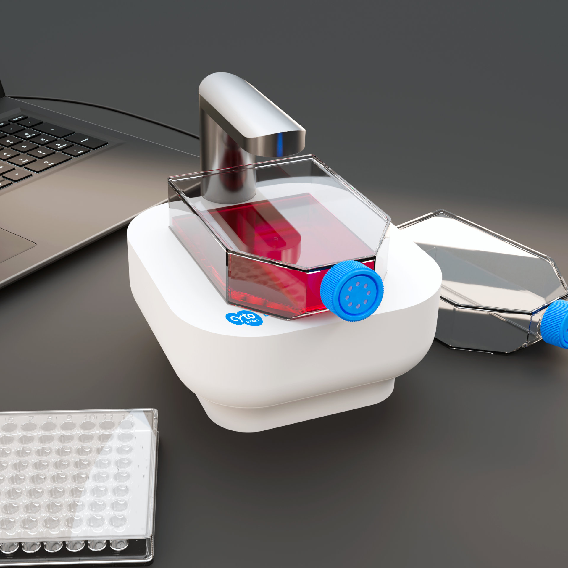
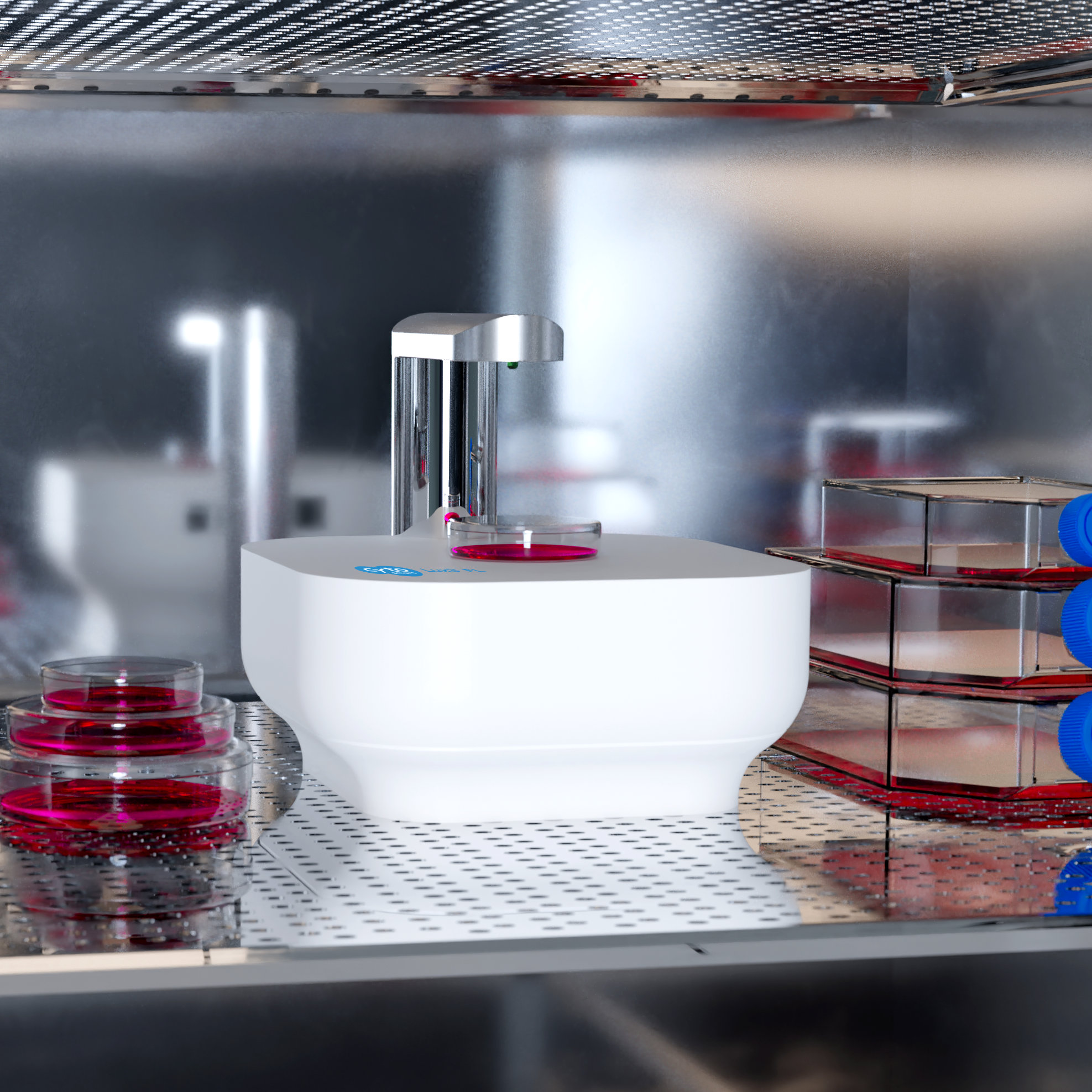
Kinetic fluorescence imaging of cellular processes requires environmental control throughout the experiment. Because of its small size, the Lux3 FL fits inside any standard incubator. This enables you to perform fluorescence live-cell imaging experiments parallel to your routine cell line passaging.
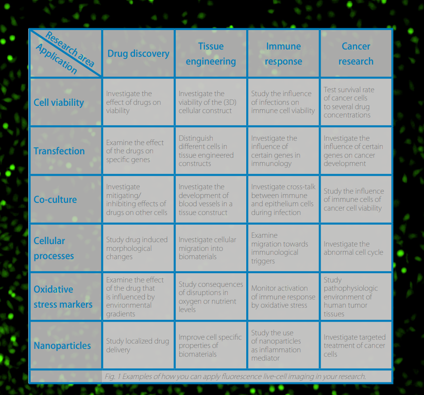
Applications
The use of fluorescence live-cell imaging has become increasingly valuable in the fields of drug discovery, tissue engineering, immunology, immunotherapy, and cancer research. With the CytoSMART Lux3 FL you have a big advantage over your colleagues and competitors. With our cloud-based solution, you have access to the following applications anywhere and anytime you need it:
- >> Cell viability
- >> Cellular processes
- >> Hypoxia / oxidative stress markers
- >> Transfection
- >> Co-culture
- >> Nanoparticles
- >> and more.
However, you are not limited to these applications or the CytoSMART image analysis software. All images and movies can be downloaded from the CytoSMART Cloud environment so you can use other (custom) image analysis algorithms if necessary.
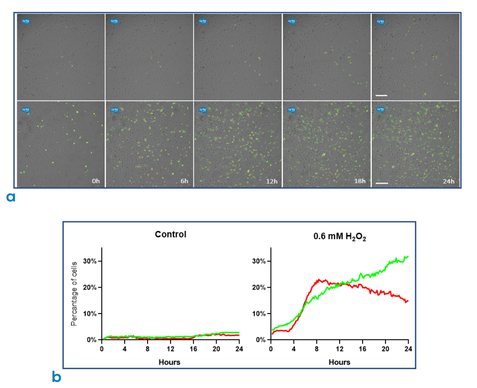
Fig 2: Fluorescence live-cell imaging of CHO-K1 cells undergoing apoptosis. a) CHO-K1 cells were either left untreated (top row) with 0.6 mM H₂O₂ to induce apoptosis (bottom row). pSIVA™-IANBD and Propidium Iodide (PI) were also added to cell culture to monitor the progression of apoptosis. Green fluorescence indicates that pSIVA™-IANBD is bound to phosphatidylserine (PS), which gets exposed during apoptosis; whereas red fluorescence indicates that the apoptotic pathway is completed. Images were captured every 10 min for a total period of 24 h. Scale bar: 200 µm. b) Automated data acquisition. Percentage of cells displaying green and red fluorescence over a period of 24 h. Left: control group; right: cells treated with 0.6 mM H₂O₂.
Live insight into cellular processes
The CytoSMART Lux3 FL fluorescence live-cell imaging microscope automatically creates time-lapse movies that contain many cellular features. The videos are made from inside the incubator without disturbing your cells and can be immediately accessed and analyzed remotely via the CytoSMART Cloud, providing real-time updates on your cell cultures and running experiments.
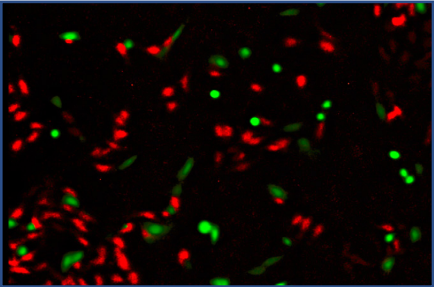
Fig 3: Co-culture of 3T3 cells stained with CellTracker Green (green) and C6 cells stained with CellTracker Orange (red).
Simultaneous analysis of multiple cell features
Using the CytoSMART Lux3 FL, you can analyze green and red fluorescent tags simultaneously with your brightfield images. This not only saves you time, but also provides tools to distinguish cells from each other or distinguish labeled structures within the cells. By tracking changes in the fluorescent signal over time, change in cell number or fluorescently-tagged cellular components can be easily identified.
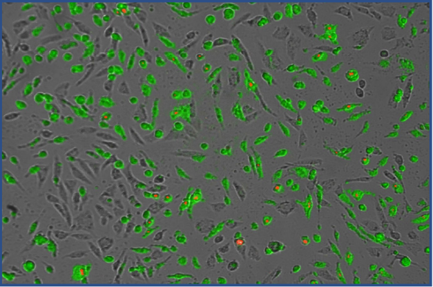
Fig 4: Expand the number of variables you can analyze in your cell culture using brightfield, green, and red fluorescent channels simultaneously.
Quantification of real-time cellular events
For certain experiments, in addition to monitoring cell cultures, it is also required to quantitatively evaluate the progression of cellular events. The CytoSMART Lux3 FL, equipped with an cell counting algorithm, can automatically calculate the number of fluorescent objects in the image and investigate how this number changes over time. What’s more, the existing software interface has become even more intuitive, allowing users to quickly and easily adjust the parameters of an image.
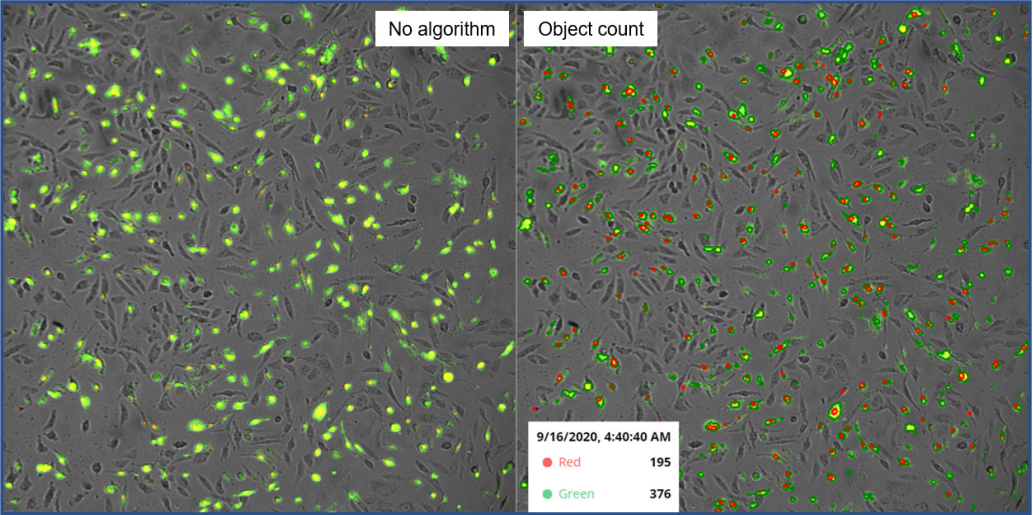
Fig 5: Analysis of apoptosis in CHO-K1 cells using pSIVA™-IANBD + Propidium Iodide (PI). Merged channels of red and green fluorescence are shown. Once applied, the Object Count algorithm (right) automatically calculates the number of red fluorescent (PI staining of nuclei) and green fluorescent (PS-bound pSIVA-IANBD) cells in the image.
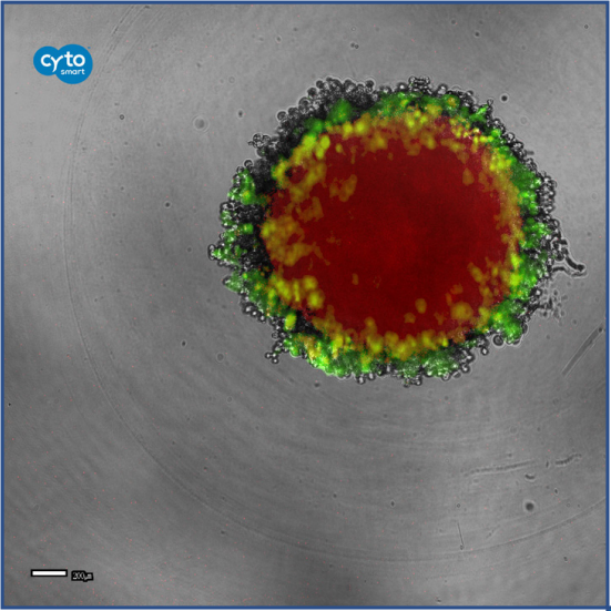
Fig 6: Proliferating cells in a 3D tumour spheroid as indicated by green fluorescence. Red fluorescence represents the drug infused in the tumour spheroid. The drug uptake and its effect on proliferation of the tumor cells are followed using the CytoSMART Lux3 FL. Image courtesy of Catarina Roma-Rodrigues, Luís R. Raposo, Prof. Pedro V. Baptista and Prof. Alexandra R. Fernandes, Faculdade de Ciências e Tecnologia, Universidade Nova de Lisboa, Portugal.
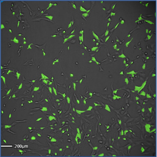
Fig 7: Overlay of green fluorescent and brightfield images of 3T3 cells transfected with CellLight Nucleus-GFP, BacMam 2.0. Images were taken 18 hours post-transfection.
Easy data storage and image analysis
The CytoSMART Lux3 FL can be set to record images at specific intervals (between 5 min - 12 h) for minutes, hours, and days. In fact, it is one of the few systems that can run for weeks. The recorded images are then sent to the CytoSMART Cloud where they are analyzed using our custom, cloud-based image analysis software. You can select the appropriate image analysis algorithm, such as confluence detection, according to the experiment you are performing.
Online connection to inspect your cell cultures
Thanks to cloud-based data storage and image analysis, you can access your recording and view the cell culture in almost real-time from anywhere on any pc, laptop, tablet or mobile phone with internet access. All the recorded data, such as images (.jpg and .tiff files), time-lapse videos (.mp4 files) or confluency data (.xlsx files) can be downloaded for further processing.

Take lab automation a step further
The CytoSMART Lux3 FL can not only be controlled via the graphical user interface of the app, but also via the CytoSMART Lux Open API. By adjusting this Python™-based open application programming interface (API) to your lab automation system, you can easily incorporate the CytoSMART Lux3 FL into your automated setup.
Plates, dishes, flasks or microfluidic chips
With the CytoSMART Lux3 FL you can image cells cultured in any culture vessel, including T-flasks, Petri dishes, well plates, culture slides, and microfluidic chips.
How to install
1. Place the CytoSMART Lux3 FL device in the incubator.
2. Connect the device’s power cable and the USB 3.0 cable to your PC or laptop.
3. Start the PC. Download the CytoSMART® Lux3 FL application.
4. Open the app and you are set to go!
Frequently Asked Questions
Q: What is the CytoSMART Lux3 FL?
A: The CytoSMART Lux3 FL is a small fluorescence live-cell imaging microscope, equipped with one brightfield and two fluorescent channels (green and red). The device allows users to track dynamic cellular processes with high specificity by taking high-quality fluorescence images to create real-time, time-lapse movies. Experimental data can be accessed and analyzed remotely via the CytoSMART Cloud.
Q: For what applications can I use the CytoSMART Lux3 FL?
A: The applications of the CytoSMART Lux3 FL include - but not limited to - monitoring cell viability, measuring transfection efficiency, investigating co-cultures, and studying dynamic cellular processes. Researchers working in the fields of drug discovery, tissue engineering, immunology, immunotherapy, and cancer research can benefit from fluorescence live-cell imaging.
Q: Is it possible to control the intensity of the CytoSMART Lux3 FL LED?
A: Yes, it is possible to set the intensity of the LED for red and green fluorescent channels according to users’ preferences.
Q: What are the software requirements?
A: The CytoSMART Lux3 FL remote functionality runs on cloud-based software. In this cloud-environment, the images and videos are stored and can be accessed using user-specific login details. Next to unlimited data storage, automated image analysis can be performed in the CytoSMART Cloud portal.
Q: Which culture flasks and dishes are compatible with the CytoSMART Lux3 FL?
A: The CytoSMART Lux3 FL allows monitoring of a wide range of different culture dishes and flasks, such as: T-flasks (T-25 up to T-250), single well, multi-well plates (6 - 384-well plates), microfluidic chips, flat tubes, Petri dishes, and slides. This is not an exhaustive list, so if your preferred equipment is not on this list please get in touch with us.
Q: What is the CytoSMART Lux3 FL magnification?
A: The CytoSMART Lux3 FL comes with a fixed 10x objective and 20x digital zoom.
Q: Can I specify the recording interval?
A: Images can be recorded at pre-defined intervals. At the start of a new experiment, you can specify the interval rate anywhere between 5 min and 12 h.
Q: How do I clean the device?
A: The device is easy to clean using lint-free wipes and ethanol (70%) or isopropyl alcohol (IPA). Do not use acetone to clean the device. Please be aware that the device cannot be autoclaved. After sterilizing with ethanol or IPA, the device can be used in a cleanroom.
Q: Which fluorescent dyes are recommended to use with the CytoSMART Lux3 FL?
A: Many different fluorescent dyes can be used, as long as the fluorescent dye’s excitation and emission spectra correspond with the fluorescent filters of the Lux3 FL (green - excitation: 452/45 nm, emission: 512/23 nm; red - excitation: 561/14 nm, emission: 630/90 nm). Some examples are green fluorescent protein (GFP) and Cell Tracker green for the green channel, and red fluorescent protein (RFP), PI, and Cell Tracker Orange for the red channel.
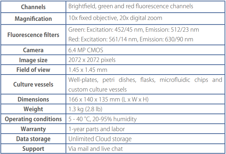
Specifications
Request quote here.