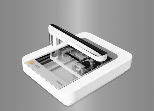
The automated live-cell imager that fits within any incubator
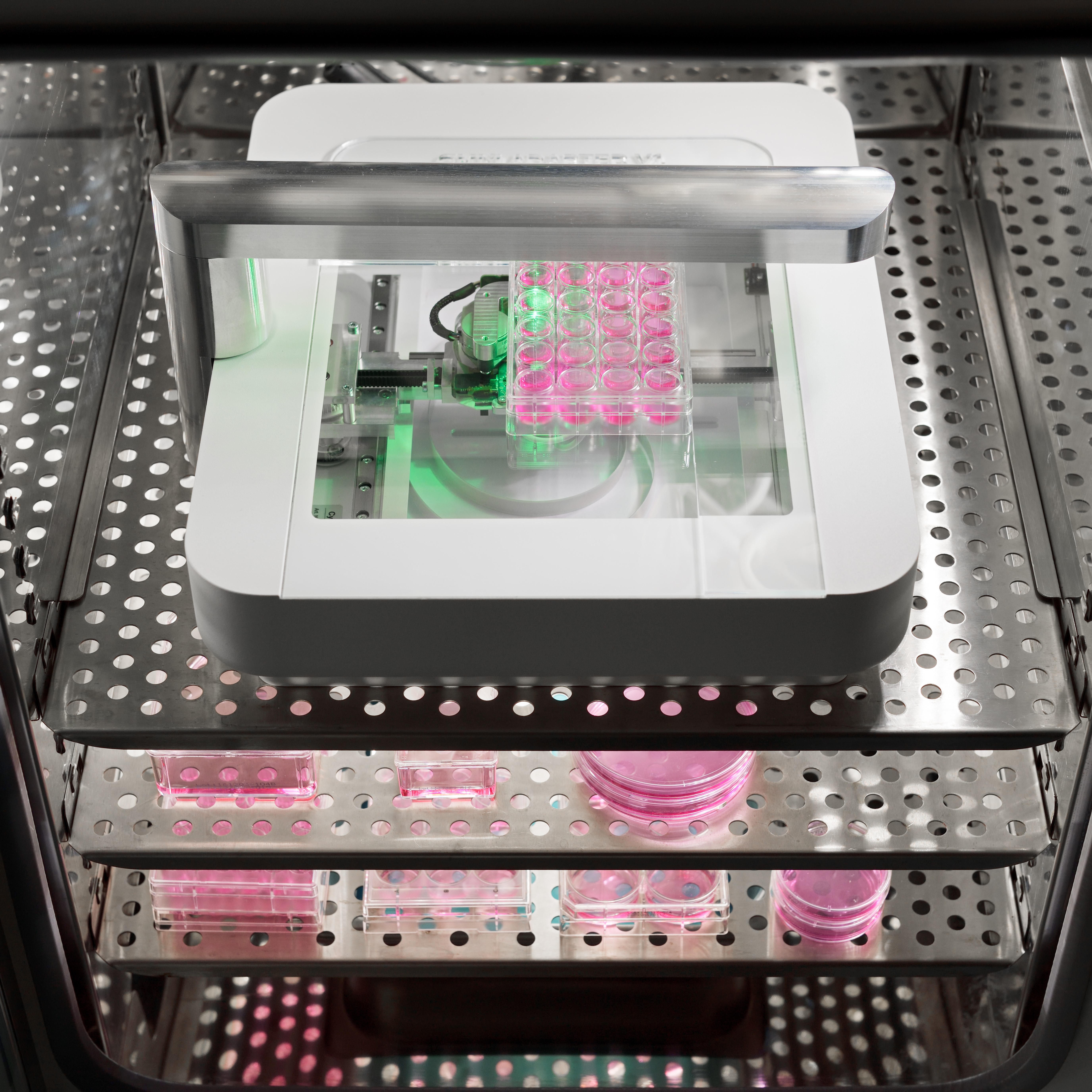
To help life science researchers improve their understanding of cellular processes, CytoSMART Technologies has developed an automated brightfield microscope that can visualize an entire surface of a cell culture vessel. It can operate from inside any standard incubator, biological safety cabinet or on a laboratory bench. Not limited to a specific type or quantity of culture vessels, the CytoSMART Omni captures cellular behavior by creating high-quality time-lapse videos for days or even weeks at a time.
The CytoSMART Omni is:
- >> Fast: 7850 images are acquired and stitched together in less than 14 minutes.
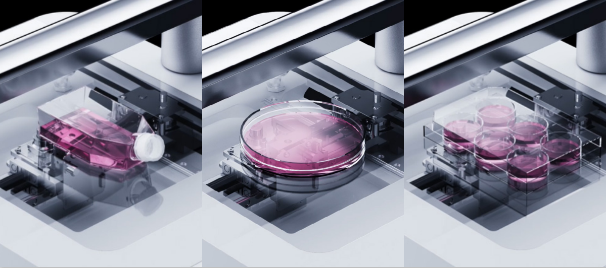
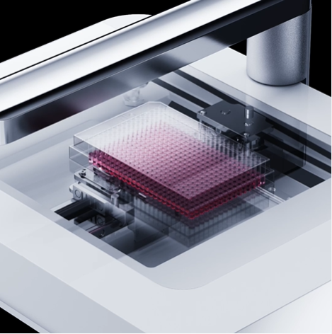
- >> Easy: Any type of transparent culture vessel can be scanned.
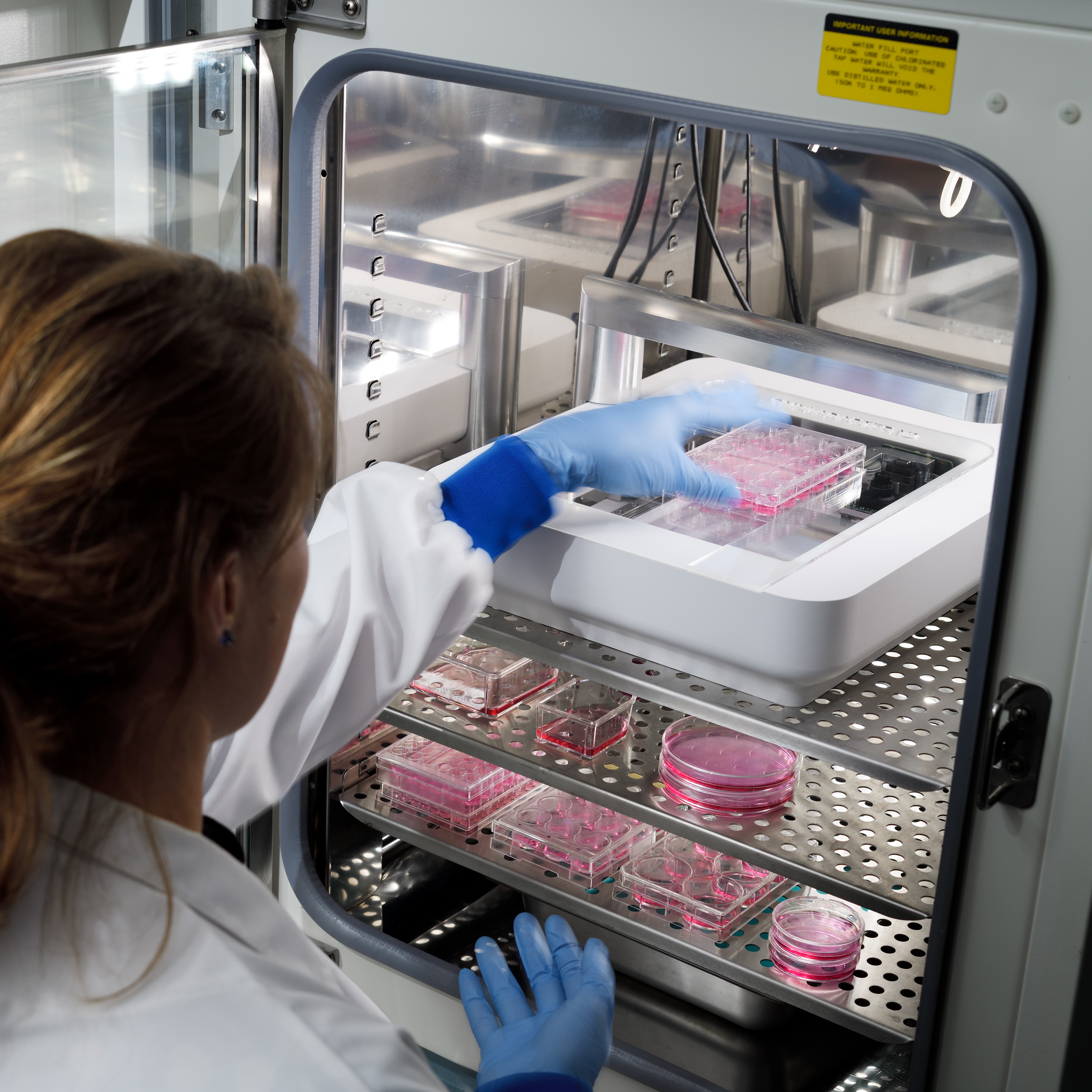
- >> Compact: Fits in any cell culture incubator.
Automated whole-well imaging with a high spatial resolution
The CytoSMART Omni, fitted with a digital 6.4 MP CMOS camera, can monitor cell cultures and study biological processes with enhanced spatial resolution, while also allowing the cells to be kept in their desired culture environment. Higher quality images coupled with robust AI-based image analysis ensure that you will generate reliable and reproducible experimental data with minimal artifacts.
Continuous imaging provides real-time insight into cellular processes
Time-lapse imaging allows you to pinpoint important events in the progression of the cell culture from experiment to experiment. The CytoSMART Omni is capable of performing kinetic assays over the course of days or even weeks. Uncover attachment and growth rates, evaluate events like cell migration and compare colony formation between wells. Data collection using video monitoring allows users to capture changes within samples and compare the rate of change between samples. With the integrated Continue Experiment feature, it is now possible to perform multiple transient screens in parallel to an ongoing long-term kinetic assay by simply pausing the running experiment.
Complete sample overview
Manual handling and cell seeding can cause a variable density distribution within culture vessels. Randomly selecting several areas of interest or tile scanning is common practice to overcome that issue. However this is time-consuming to set up and to post-process, and can lead to inconclusive data. The CytoSMART Omni automatically scans complete well surface areas and instantly stitches these images to give users a complete overview of cell coverage (Fig. 1).
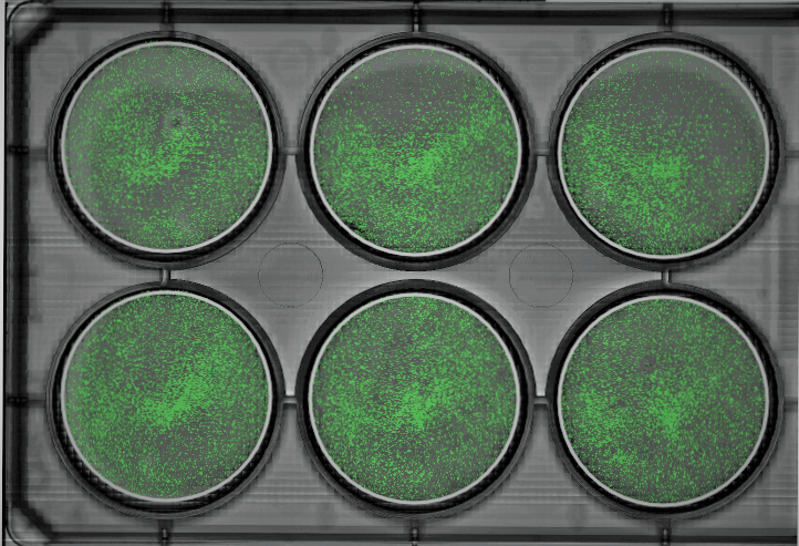
Fig 1: Scan of a 6 well-plate, with the confluency algorithm overlay, showing an inequal distribution of the cells through each well.
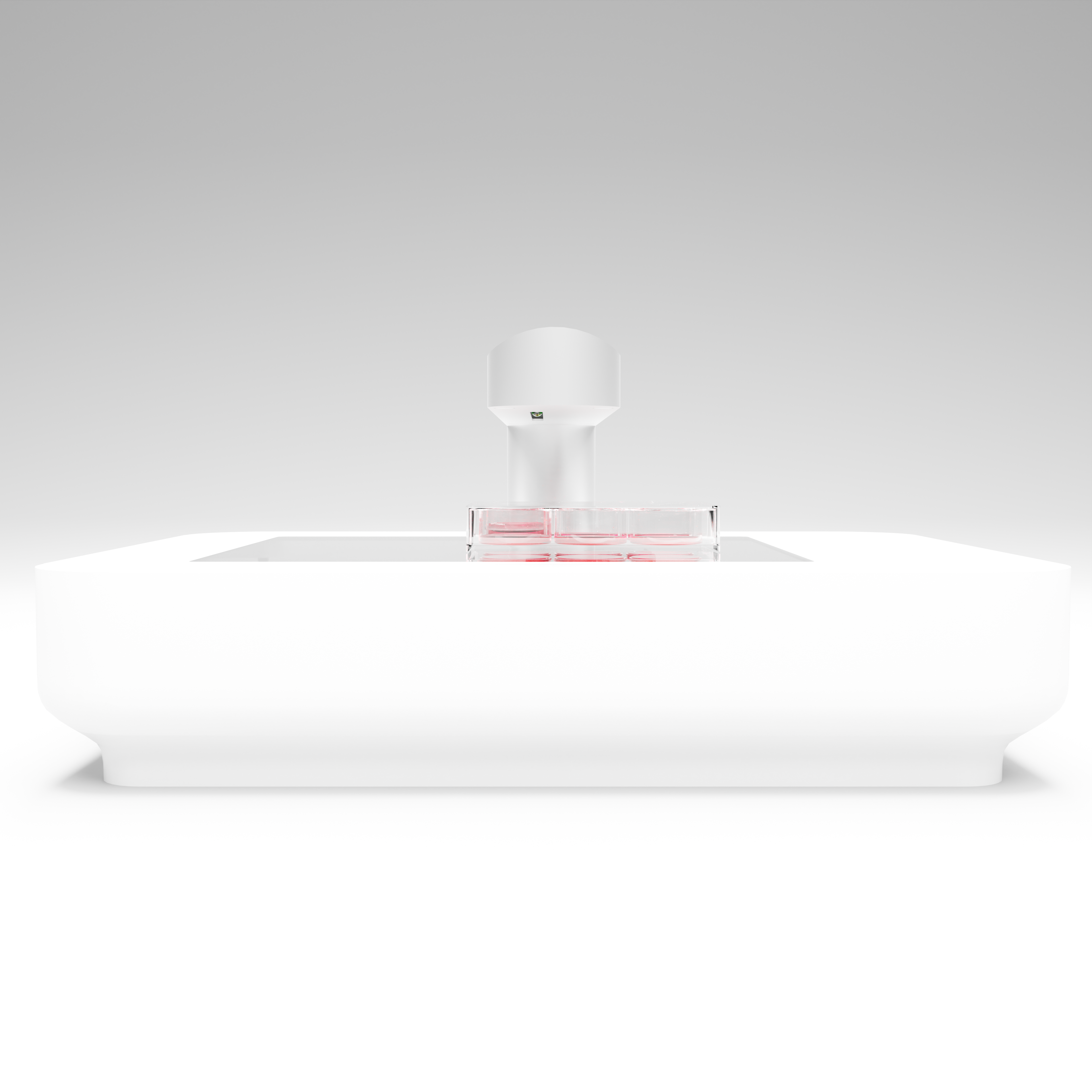
Compatible with any transparent culture vessel
Cell-based assays and subculturing all benefit from standardization. Choosing the right vessel and going through the process of understanding how cells behave in said vessel is vital for reproducibility. The CytoSMART Omni is highly flexible because of its open and flat design, and therefore compatible with any transparent vessel that is lower than 55 mm. 6 – 384 well plates are automatically recognized by AI and deep learning algorithms. Other culture vessels, such as T25 – T225, Petri dishes, triple flasks, HYPERFlasks and microfluidic chips are imaged in full and analysed with custom regions of interest.
Easy access. Anywhere. Anytime.
Thanks to cloud-based data storage and image analysis, you can access your CytoSMART Omni experiments in almost real-time from anywhere on any pc, laptop, tablet or mobile phone with internet access. In case you have set a notification, our email alerts will keep you up-to-date on confluence levels or long-term temperature drops. Get rid of hard drives or USB-drives for data transfer at the end of your experiment: just easily connect to the Cloud via any internet-connected device, and view, adjust and analyze the data accordingly. Besides the advantage that any adjustment in the Cloud is processed in retroactivity and in real-time, it also allows for unlimited data storage. Share collective experiments with colleagues while retaining tight control of the data analysis and output. All the recorded data such as images (.jpg files), time-lapse video (.mp4 files) or image analysis data (.xlsx files) can be downloaded for further processing.
Applications
The large scanning area of the CytoSMART Omni can provide complete sample information in cancer research, drug discovery, biomaterial research, fundamental cell and tissue research, tissue engineering, and many other research areas.
With the integrated cloud-based image analysis, data evaluation and quantification are just a few clicks away. The CytoSMART Omni provides you access to the following applications:
- >> Confluence measurement to observe cellular proliferation and cell death over time.
- >> Cell culture quality control to get immediate insight into the influence of different culture conditions on cell health (Fig. 2)
- >> Cell confluence in cytotoxicity assays (Fig. 3)
- >> Collective cell migration in cell removal assays (Fig. 4), cell exclusion assays (Fig. 5) and cell outgrowth assays, by means of area closure and closure speed
- >> Single colony area, size and circularity in clonogenic assays, to monitor cancer cell or stem cell colony growth over prolonged periods of time (Fig. 6)
- >> and more.
However, if the available image analysis algorithms are not sufficient for your needs, the raw images can be analysed using any commercially available image analysis program.
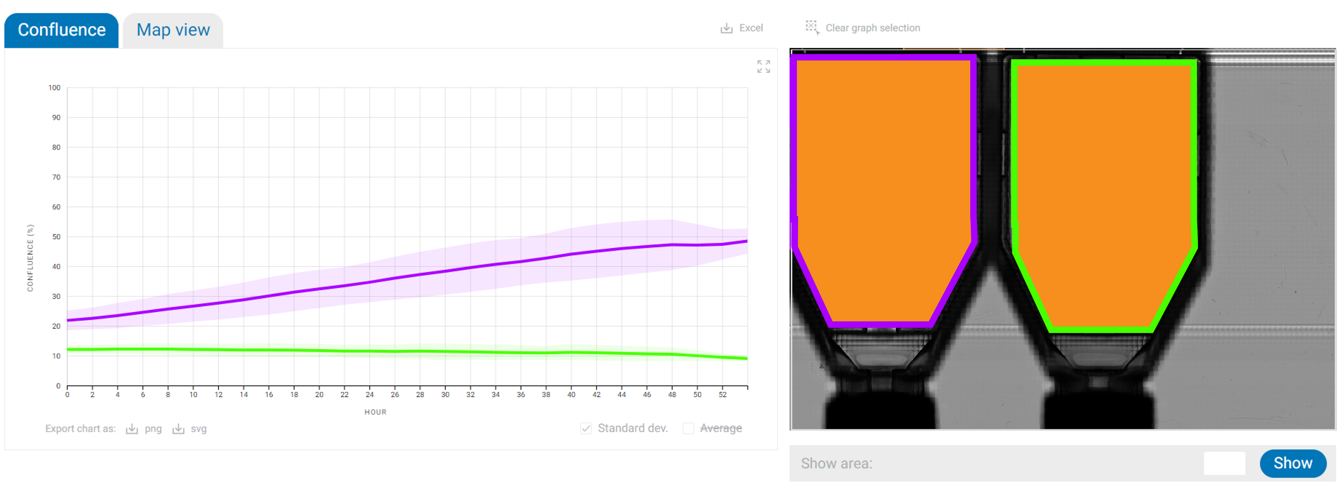
Fig 3: Confluence analysis for easy and direct observation and understanding of cell proliferation rates. Left flask: human dermal fibroblasts cultured in keratinocyte growth medium. Right flask: human dermal fibroblasts cultured in 75% keratinocyte growth medium and 25% fibroblast growth medium.
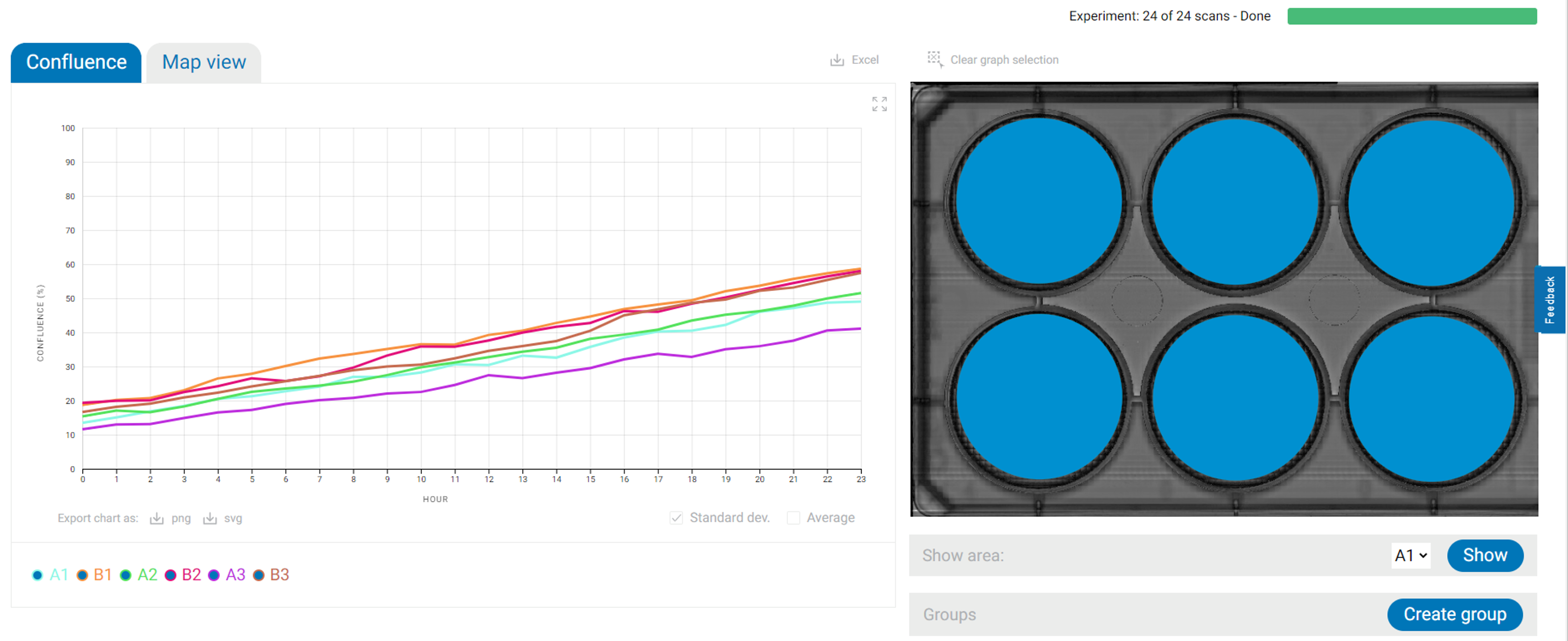
Fig 3: As cell confluence is directly related to cell death, effortless and straightforward cytotoxicity data is easily obtained over time. Graph: normalized confluency of C6 cells treated with 12 different concentrations of Paclitaxel (mean ± standard deviation; n=12).
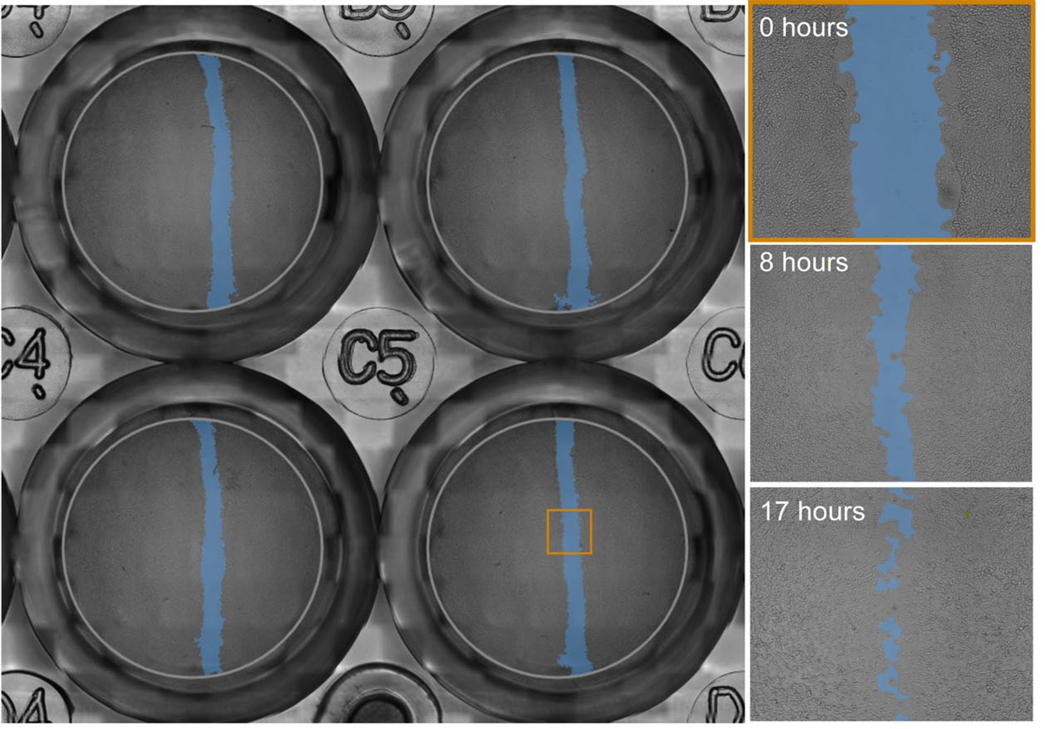
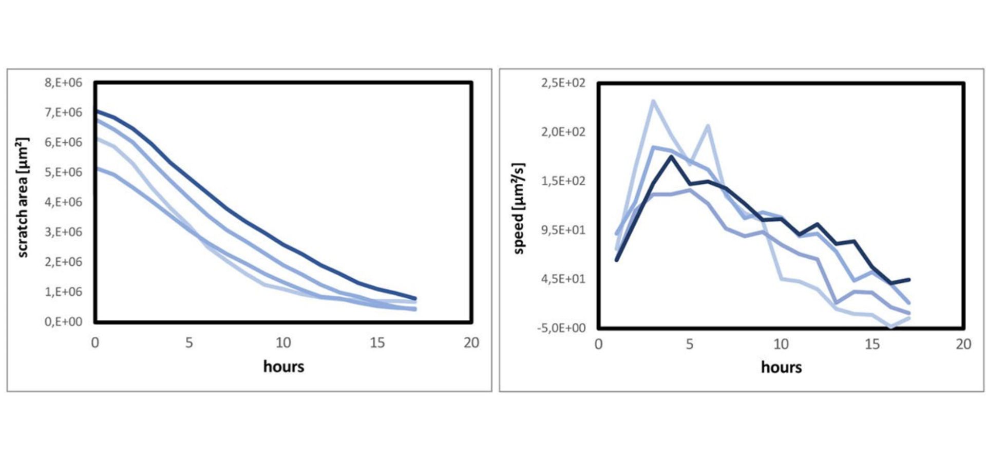
Fig 4: Upscale cell migration experiments by receiving full well plate data automatically. The image analysis provides information on gap surface closure and closure speed over time from a whole well to cell level detail.
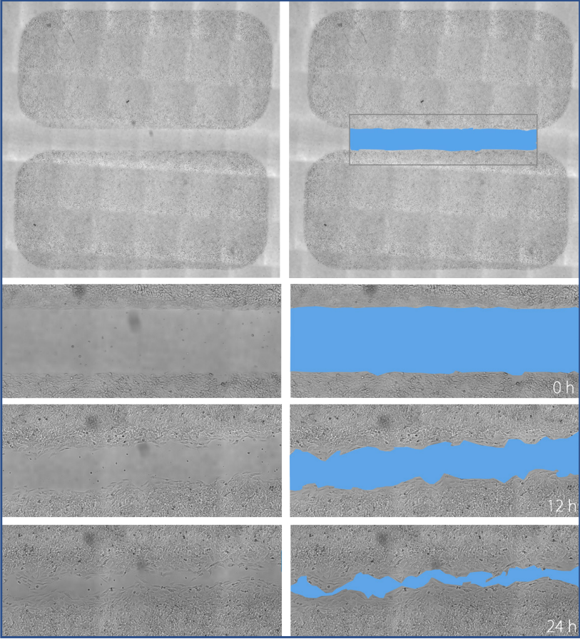
Fig 5: Inserts can be ideal for quick and reproducible wound healing assays. Regions of interest (gray box) are quickly created, ensuring that only interesting areas are displayed. (Courtesy of Hanan Osman-Ponchet, PKDERM).
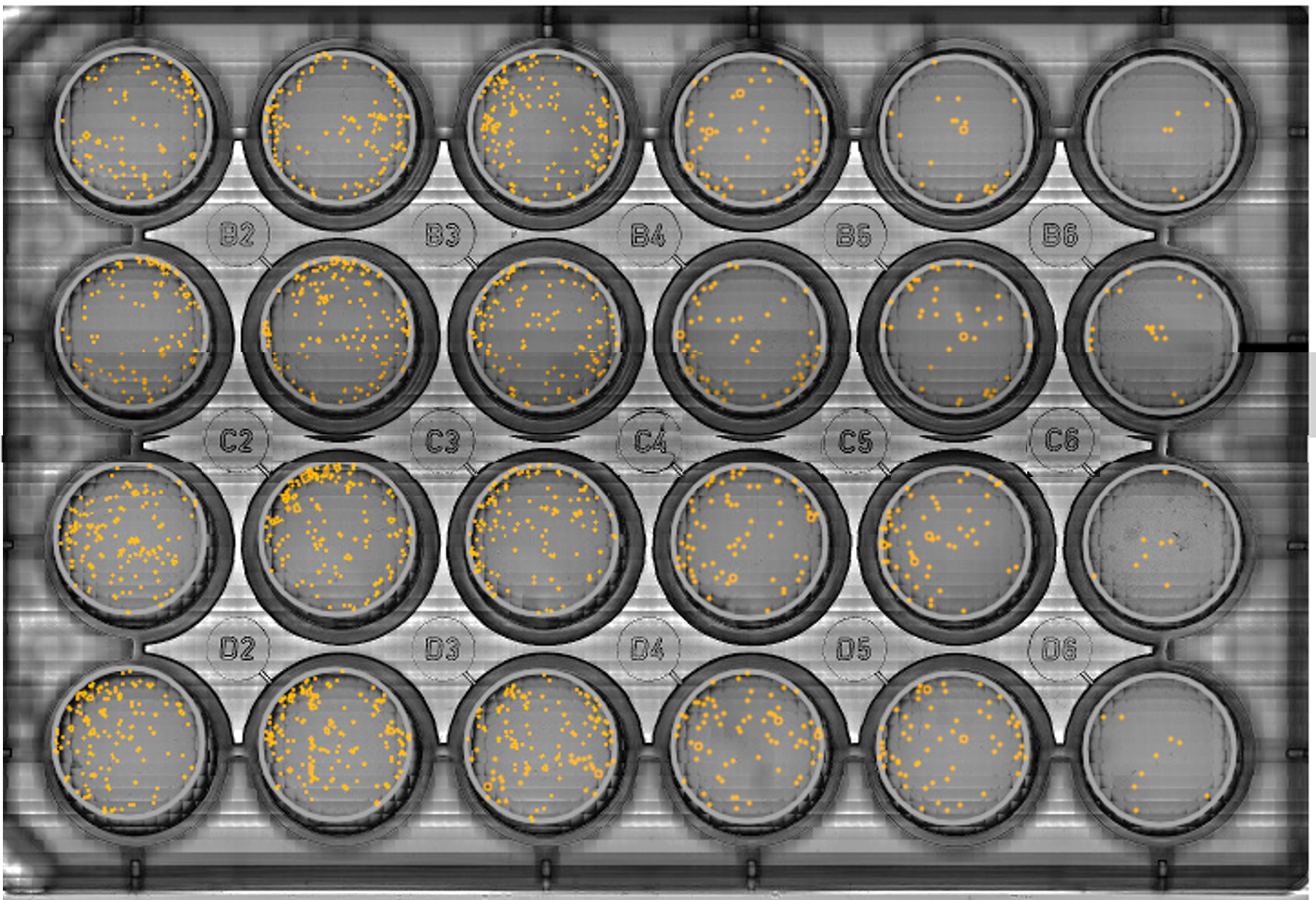
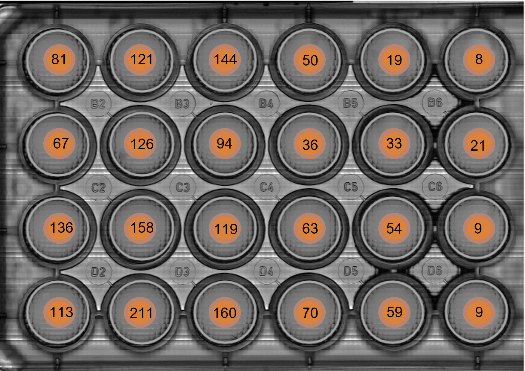
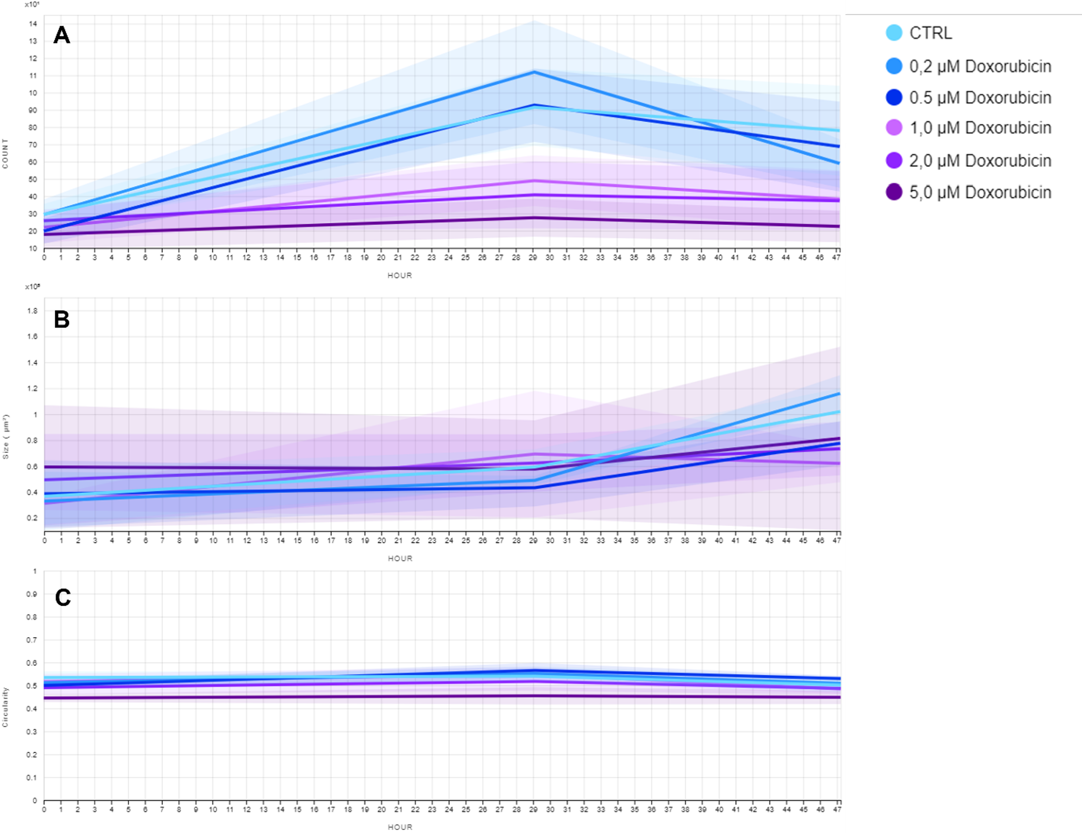
Fig 6: Follow colony formation over time and obtain qualititave and quantitative data in parallel, to draw conclusions sooner. CHO-K1 cells were treated with different concentrations of Doxorubicin. Using the analysis algorithm for clonogenic assays, data analysis was accelarated by evaluating single colony area, size and circularity, while biased and subjective interpretation of the results was reduced. a) Overview of the detected colony outlines. b) Number of counted colonies per well. c) Average and standard deviation of colony count (A), size (B) and circularity (C) over time per Doxorubicin concentration.
Frequently Asked Questions
Q: How does the CytoSMART Omni work?
A: The device takes 7850 images in an area of 86 x 124 mm, that are uploaded to the CytoSMART Cloud. There, they are combined into one scan and subsequently analyzed using our custom image analysis algorithms.
Q: Can I specify the recording interval?
A: You can specify the interval rate between 1h - 24 h or choose to perform a single scan.
Q: What is the magnification of the CytoSMART Omni?
A: The magnification is equal to the magnification of a microscope with a 10× objective.
Q: What type of image analysis algorithms can I use?
A: Confluence, collective cell migration and colony formation analysis are available in the Cloud. All images and videos can also be downloaded for personal image analysis.
Q: Can the CytoSMART Omni be used inside a cell culture incubator?
A: Yes, the CytoSMART Omni is designed to be used inside a cell culture incubator. Its hardware and electronics can operate at 5 - 40 °C and 20 - 95% humidity.
Q: Do I need to label my cells in order to perform image analysis?
A: No, our image analysis algorithms are designed to evaluate unlabeled cells, so you do not have to add (toxic) dyes to your cells. This enables to non-invasive analysis of your cells.
Q: Is a computer required to operate the CytoSMART® Omni?
A: Yes, the device can only be used with a Windows-based computer with a USB3.0 port (which can also be purchased at CytoSMART). A WiFi or wired ethernet connection is necessary to be able to connect to the CytoSMART® Cloud for data storage and analysis.
Q: Which culture vessels are compatible with the CytoSMART Omni?
A: Any culture vessel lower than 55 mm (height of the light arc) can be scanned. However, the size of the scan is limited to 86 x 124 mm, which fits a regular well plate or T175 flask. In case you would like to image larger vessels, you cannot capture the entire surface.
Q: Why is the CytoSMART Omni a cloud-based device?
A: The scans are uploaded to the CytoSMART® Cloud because of the vast amount of data that is created with the CytoSMART Omni. In order to store and analyze the gigabytes of data, you would need a high-end local computer which costs a lot more compared to cloud-based storage and analysis. Furthermore, the CytoSMART Cloud is powered by the Microsoft Azure, which is one of the most secure cloud-platforms. This ensures safe storage of your data and allows you to retain tight control over data sharing.
Q: How do I clean the CytoSMART Omni?
A: Clean the device using lint-free wipes and 70% ethanol or isopropyl alcohol (IPA). Do not use acetone to clean the device. The device cannot be autoclaved.
Q: Can the CytoSMART Omni be used in a cleanroom?
A: Yes, after sterilizing with 70% ethanol or IPA, the device can be used in a cleanroom.
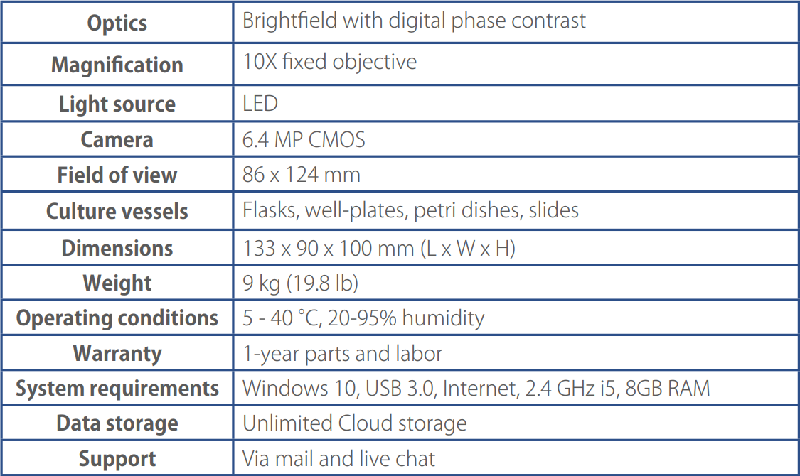
Specifications
Ordering information
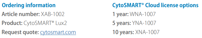
Request quote here.