Transfection and transduction are processes used to introduce foreign nucleic acids into eukaryotic cells, resulting in modification of the host cell genome. Transduction uses viral methods to transfer the genetic material, while transfection uses non-viral means. Transfection or transduction efficiency, which can be assessed using a variety of approaches, reflects the proportion of cells in a sample that acquired a foreign element. Discover how our live-cell imaging and cell counting platforms for monitoring and quantifying the time course of transfection provide valuable, time-dependent insight into the process.
-
Monitor and quantify the uptake and expression of foreign elements by transfected cells>
-
Optimize transfection protocols by testing different transfection conditions>
-
Application note: Analysis of BacMam Transduced HeLa Cells with High-Throughput Live-Cell Imaging>
Transfection is a powerful tool for studying cell physiology and complex molecular mechanisms of diseases, as it can shed more light on gene regulation and protein function. Direct visualization and quantification of the uptake of fluorescently-labelled constructs is enabled by the advanced imaging capabilities and the machine-learning algorithms of our live-cell imaging systems.
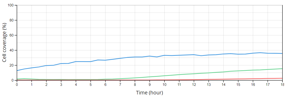
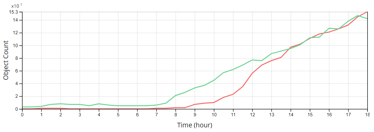
HepG2 cells were transduced with the BacMam system and imaged using the Lux3 FL. Seven hours post-transfection, a rapid increase in BacMam-Actin-GFP and BacMam-Nucleus-RFP can be observed, as detected by the Object Count algorithm.
Optimization of transfection conditions is a critical step in any transfection protocol. Variables, such as seeding density, nucleic acid quantity, and transfection time, among many others, can be optimized to improve transfection efficiency. Our devices can be used to optimize multiple steps of a transfection procedure. A comparison of different transfection parameters (seeding density and BacMam vector concentration) was performed using the Omni FL.

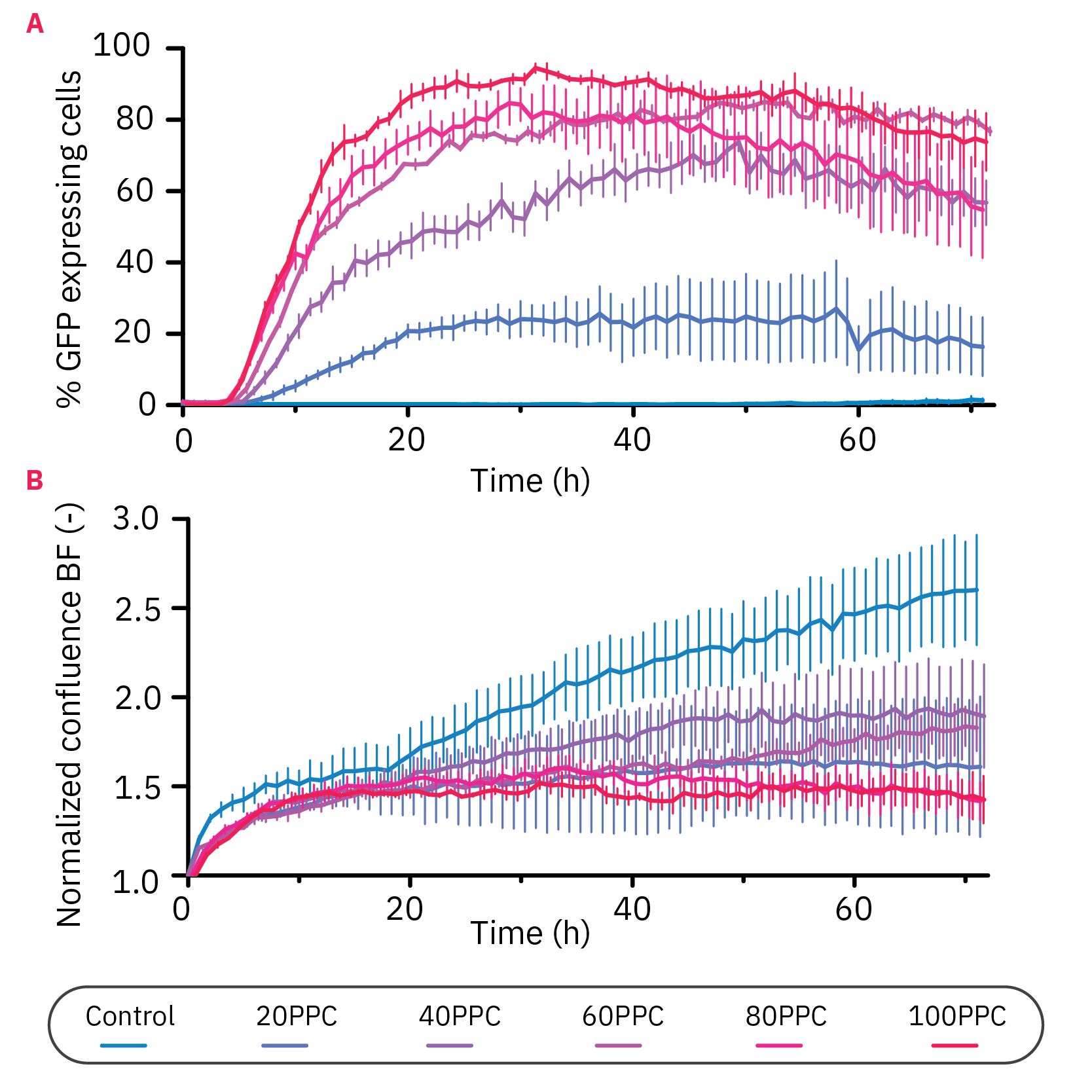
The transfection efficiency of Nucleus-GFP construct in HeLa cells is affected by the seeding density, vector concentration, and incubation time.