Image
Featured Application
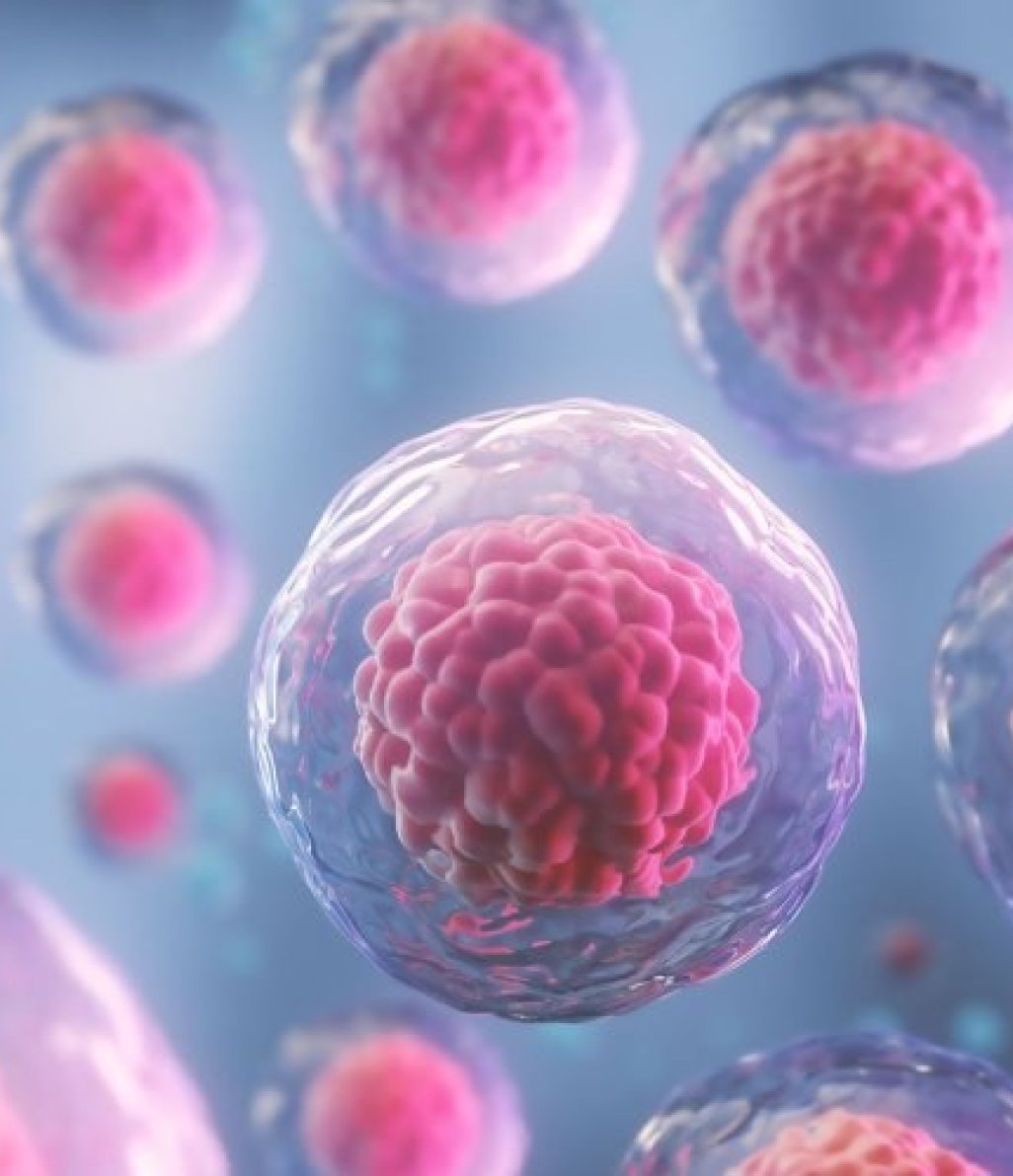
Cell Counting
Measure cell concentration, size, and viability for single cell or organoid cultures.
Learn More
→
Cancer is a complex disease that results from a series of successive genetic mutations, leading to defective cell functions and proliferation. The development of novel scientific techniques, alongside the optimization of already existing approaches, drives the advancement of cancer research.
Live-cell microscopy has become an indispensable tool for improving our understanding of cancer pathogenesis. Whether directly visualizing the transient interactions between immune and cancer cells or examining complex biological processes, such as metastasis, programmed cell death and tumor angiogenesis, time-lapse imaging can provide unique insights.